Medicine and forensics
Medicine has made enormous advancements in recent years. A key element of this development is the use of the latest materials, which were unimaginable until recently. Developments in this field require powerful analytical technology. Furthermore, the production process necessitates very high standards in terms of quality assurance. For medicine and medical technology, JEOL supplies innovative solutions across a wide range of segments.
In forensics, specialist laboratories and authorities around the globe rely on JEOL systems. Thanks to extremely powerful and highly sensitive measuring instruments, it is possible to visualise even the smallest of traces and thus clarify the circumstances and issues involved in forensic cases.
Detailed solutions
- 3D reconstruction of biological micro- and nanostructures
3D reconstruction of biological micro- and nanostructures
The resolution of conventional optical microscopes and x-ray tomography systems is insufficient to understand the structural information of biological samples on a micro- and nanoscale. With JEOL electron microscopes it is possible to image and three-dimensionally reconstruct not only the smallest of biological structures such as viruses, but also microscale objects such as synapses or cells.
Product groups
Solutions
Medicine and forensics
 Reconstruction of a bacteriophage (left) and synapses (right)
Reconstruction of a bacteriophage (left) and synapses (right)
Source data: Journal of Structural Biology 177 (2012) 589–601 and JEOL News 51
- Biocide analysis
Biocide analysis
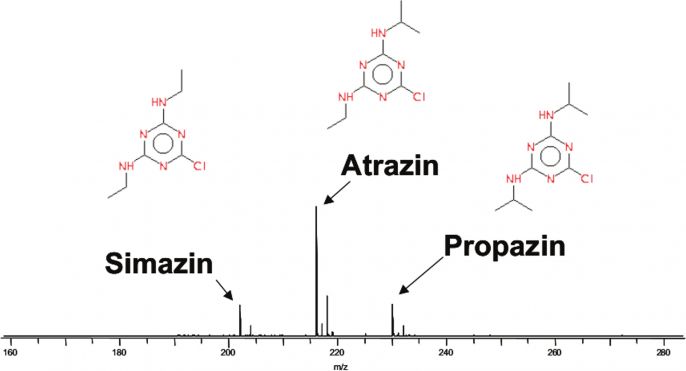
These days, biocides are used extensively for pest control in agriculture. The quantitative determination of the toxic residues of the applied pesticides is of vital importance for the authorisation of agricultural products. JEOL high-resolution analytical instrument are capable of detecting biocides quickly and simply, even with the smallest of concentrations.
Product groups
Solutions
Medicine and forensics
 Mass spectrum of various herbicides
Mass spectrum of various herbicides
Source data: JEOL Ltd. / DART Application Notes, page 57
- Characterising biofunctional surfaces
Characterising biofunctional surfaces
The surface of lotus leaves can serve as a model for self-cleaning surfaces. The exact characterisation of these surfaces is vital in order to be able to recreate them. The surface of lotus leaves comprises small wax tubes that can be easily destroyed during examination with an electron beam. JEOL therefore offers tailor-made solutions that thermally stabilise the samples in a controlled manner and thus prevent them from being destroyed by the observation.
Product groups
Solutions
 Surface of a lotus petal. The wax tubes have a diameter of approx. 50 nm
Surface of a lotus petal. The wax tubes have a diameter of approx. 50 nm
Source data: JEOL (Germany) GmbH
- Characterising biomolecules #2
Characterising biomolecules #2
Cryo electron microscopy and the analysis of protein structures has undergone drastic changes since the last years. This technique was awarded the Nobel Prize in Chemistry 2017 (https://www.nobelprize.org/nobel_prizes/chemistry/laureates/2017/advanced-chemistryprize2017.pdf).
With the introduction of fast and highly sensitive camera techniques as well as dedicated, automated electron microscopes resolutions of 2 Ångström and lower could be achieved under near to native conditions. This single particle analysis method (SPA) allows to efficiently determine the structure of non crystalline proteins in order to design new drugs or to resolve the fundamental functions of biochemical or molecular biological processes.Product groups
Solutions
Medicine and forensics
 GroEL protein at 40k x magnification (pixel size 0.12 nm), detector: K2 summit, instrument: CRYO ARMTM200, Schottky 200kV, inset: reduced live FFT, zero-loss image with 20eV slit width
GroEL protein at 40k x magnification (pixel size 0.12 nm), detector: K2 summit, instrument: CRYO ARMTM200, Schottky 200kV, inset: reduced live FFT, zero-loss image with 20eV slit width
Source data: JEOL Ltd., University Osaka, Prof. Namba
- Characterising biomolecules
Characterising biomolecules
DNA origami is a new technology in synthetic biology, or rather the disciplines of biochemistry and biophysics. It involves DNA molecules being turned into random two- and three-dimensional nanoforms. These synthetically folded DNA molecules are used as e.g. future biocompatible carriers for active substances or to produce nanomachines or nanorobots.
Product groups
Solutions
Medicine and forensics
 Negatively contrasted TEM image of synthesised DNA molecules
Negatively contrasted TEM image of synthesised DNA molecules
Source data: JEOL (Germany) GmbH, Technical University of Munich, Prof. Dietz and Klaus Wagenbauer.
- Characterising cryostabilised samples
Characterising cryostabilised samples
Specific preparation methods must be used for the high-resolution imaging and analysis of biological samples in an electron microscope. Food stuffs and their constituents in particular can only be imaged artifact-free through active cooling. JEOL electron microscopes are therefore prepared as standard for the installation of cryogenic systems so that sensitive samples can be prepared externally, transferred in a cooled state and subsequently examined in a cryogenic mode of operation.
Product groups
Solutions
Medicine and forensics
 Electron microscope image of powdered milk
Electron microscope image of powdered milk
Source data: JEOL (Germany) GmbH, DIL Quakenbrück
- Element analysis
Element analysis
Magnetotactic bacteria orientate themselves along the Earth's magnetic field with the help of magnetite particles enveloped in a membrane. These organisms serve as a model for examining the complex processes of biomineralisation. Entities known as magnetosomes (comprising magnetite crystal and membrane) are also tested as carriers for active substances and for new forms of therapy in medicine (hyperthermia therapy). JEOL supplies combined and fully automated solutions for the high-resolution elementary classification of the individual constituents.
Product groups
Solutions
Medicine and forensics
 Element analysis (red rectangle) at 120 kV in S(TEM) on magnetotactic bacteria, magnetite chains (green)
Element analysis (red rectangle) at 120 kV in S(TEM) on magnetotactic bacteria, magnetite chains (green)
Source data: JEOL (Germany) GmbH
- Imaging moist samples
Imaging moist samples
To achieve the high-resolution imaging and analytics of biological samples, it is often necessary to examine the sample in its native state. Thanks to the patented JEOL Aqua Cover, it is even possible to image moist or hydrated samples in a scanning electron microscope at low pressure.
Product groups
Solutions
 Image of a water droplet on the surface of a rose petal
Image of a water droplet on the surface of a rose petal
Source data: JEOL Ltd. (Aqua Cover presentation)
- Pinpointing fluorescently labelled proteins
Pinpointing fluorescently labelled proteins
The linking of light microscopy signals and electron-optical details allows, among other things, conclusions to be drawn regarding the exact location of proteins in specific sections of tissues. JEOL manufactures intuitive, multi-system complete solutions for combining fluorescence and high-resolution electron microscopy.
Product groups
Solutions
Medicine and forensics
 Thin section of a zebra fish: correlatively superimposed electron and fluorescence microscopic images
Thin section of a zebra fish: correlatively superimposed electron and fluorescence microscopic images
Source data: JEOL (Germany) GmbH, Centre for Regenerative Therapies, TU Dresden.
- Preparation and characterisation of fragile structures
Preparation and characterisation of fragile structures
The colours of a butterfly wing are created by pigment or structural colours. Due to the delicate surface of the wing, it is mechanically impossible to prepare a cross-section. With JEOL preparation systems and scanning electron microscopes, even fragile, organic structures can become accessible and visible.
Product groups
Solutions
Medicine and forensics
 Image of a cross-section through a butterfly wing (Morpho)
Image of a cross-section through a butterfly wing (Morpho)
Source data: JEOL Ltd., Cross Section Polisher brochure
- Tissue diagnostics
Tissue diagnostics
As part of a biopsy, tissue is removed and subsequently examined under the microscope. Labels can also be used to pinpoint the location of active substances to be examined. JEOL not only supplies specially developed, automated systems for simple and high-constrast imaging and high-resolution and ultrastructural diagnostics. With highly sensitive JEOL EDX detectors, it is also possible to pinpoint the location of the labels used.
Product groups
Solutions
Medicine and forensics
 TEM image of a tissue section
TEM image of a tissue section
Source: JEOL (Germany) GmbH, Demo Friedrich Baur Institut in Munich
