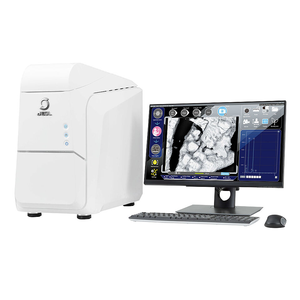
JEOL JCM-7000 Table-top Scanning Electron Microscope
JEOL JCM-7000 Table-top Scanning Electron Microscope
Never has SEM imaging been so easy: the new JCM-7000 incorporates straightforward sample navigation and advanced auto functions making it the ideal tool for users at any skill level to obtain outstanding SEM images and elemental analysis results in minutes. It unites SEM images, reconstructed 3D surfaces and EDS elemental analyses in real-time within an intuitive user interface. In addition, the mobile all-round talent JCM-7000 features a large sample chamber, high resolution and a low-vacuum mode for challenging samples.
Features
- “Live 3D”: real-time SEM image and reconstructed 3D surface for simultaneous acquisition of topographic depth information and surface fine structures.
- Full-featured electron optics for magnifications up to x100,000.
- Straightforward emitter change provided by pre-centered tungsten filaments.
- Automatic condition setting based on sample type and application ensures high quality results and enhances productivity.
- JEOL ZeroMag for seamless transition between light optical and SEM imaging dramatically improves handling and through-put.
- Fully integrated JEOL EDS system including live EDS for maximum ease of use and comprehensive report generation.
- Automated acquisition of SEM images and elemental distribution maps resulting in high resolution montages.
- Challenging samples can be investigated in low-vacuum mode with the push of a button.
- Large chamber for samples of up to 80 mm diameter and 50 mm height.
- Simple installation – an electric socket and you are ready to go!
- Compact design for mobile applications.
Specifications
Magnification | ×10 to 100,000 (Reference: Polaroid) |
Accelerating voltage | 5 kV / 10 kV / 15 kV |
Imaging | Secondary electron detector (SE) and backscattered electron detector (BSE) in high vacuum mode, backscattered electron detector (BSE) in low vacuum mode |
Electron Gun | Tungsten source (pre-centered filaments) |
Specimen stage | 2 axes motorized (X: 40 mm, Y: 40 mm) |
Specimen size | Max. diameter: 80 mm, max. height: 50 mm |
Operation | Keyboard, mouse |
Options
- Energy-dispersive X-ray spectroscopy (EDS)
- Motorized tilt/rotation holder
- Color stage navigation system (SNS)
- Fully automated particle analysis software

Please note:
Subject to technical changes; errors excepted. All brand names that appear in the text are registered trademarks of the manufacturers.